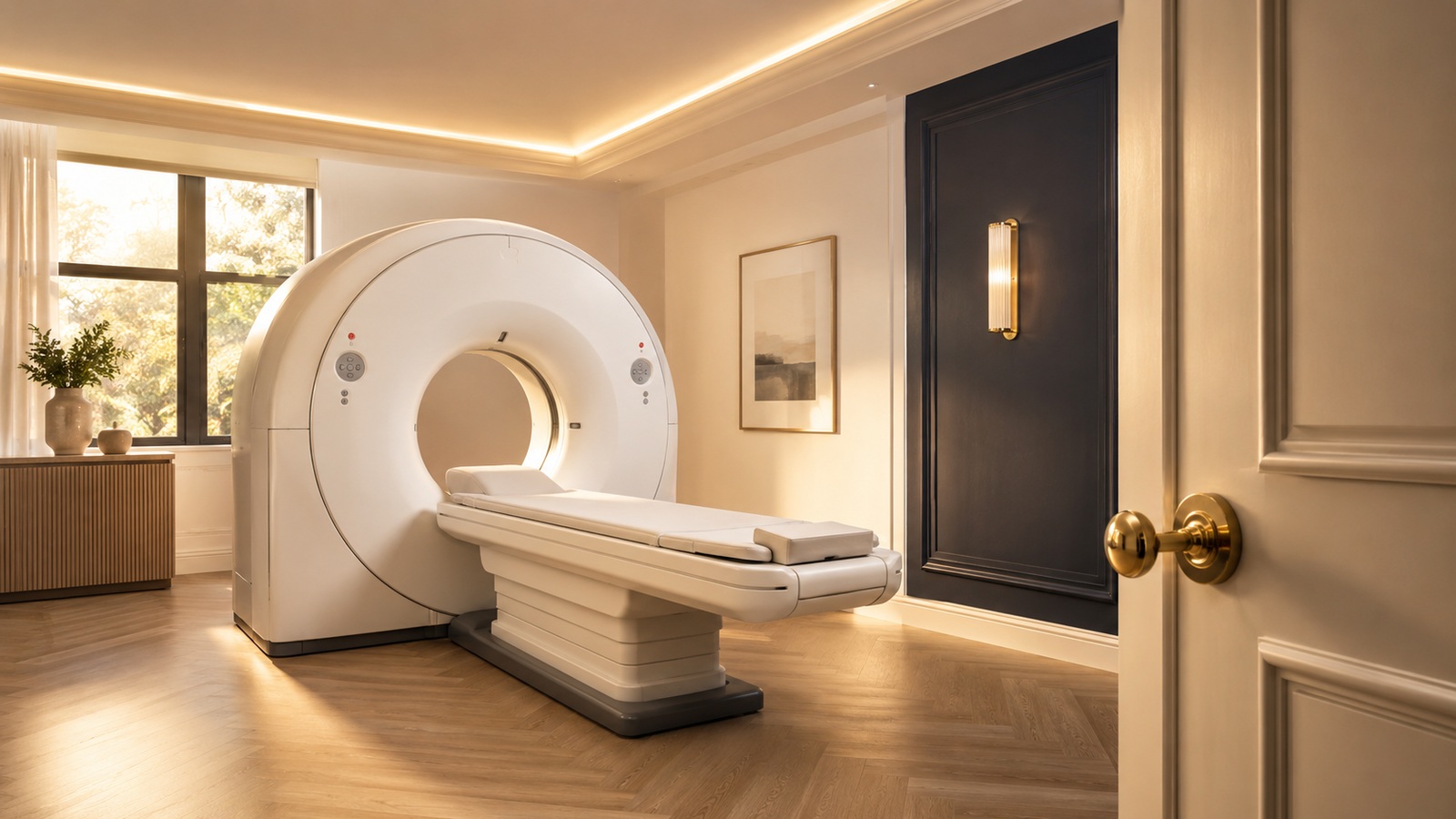
The pathway, in order
For chest pain that could be angina, UK national guidance (NICE) recommends CT coronary angiography as the first-line investigation. If its result is unclear or the question becomes how much a narrowing actually restricts blood flow, functional imaging comes second. The invasive angiogram — a catheter passed to the heart — is kept for when its answer will change treatment [1][2].
What the CT shows
A CT coronary angiogram photographs the heart's arteries themselves, from the outside in, without a catheter: a scan with contrast dye through a vein in the arm [1][4]. It shows whether the arteries are clear, streaked with early plaque, or significantly narrowed — and a clean result is powerful, because it makes a coronary cause of the pain very unlikely and can close the question in one visit [1][2].
Why CT first makes sense
Most people investigated for stable chest pain turn out not to have a dangerous narrowing. A first test that is non-invasive, quick, and excellent at ruling coronary disease out spares most patients an invasive procedure they never needed — while still catching the disease it exists to find [1][2]. When the CT does show significant disease, everything that follows — medication, further tests, or treatment of the narrowing — starts from an anatomical map that has already been drawn [1][2].
What happens after the scan
Three outcomes cover most cases [1][2]:
- Clear arteries — the pain is very unlikely to be coronary, and attention turns to other causes and to your risk factors.
- Plaque without a tight narrowing — the priority becomes prevention: blood pressure, cholesterol and lifestyle, treated seriously.
- A significant narrowing — the findings go to a cardiologist, and any decision about treating it is made with you, on the evidence.
A note on the day itself
The scan is quick and painless; a medicine to steady the heart rate is sometimes given first so the pictures are sharp, and the radiation dose of modern scanners is low [2][4]. The result is read by a specialist in cardiac imaging — the picture is only as good as the reading of it.