What is a cardiac MRI?
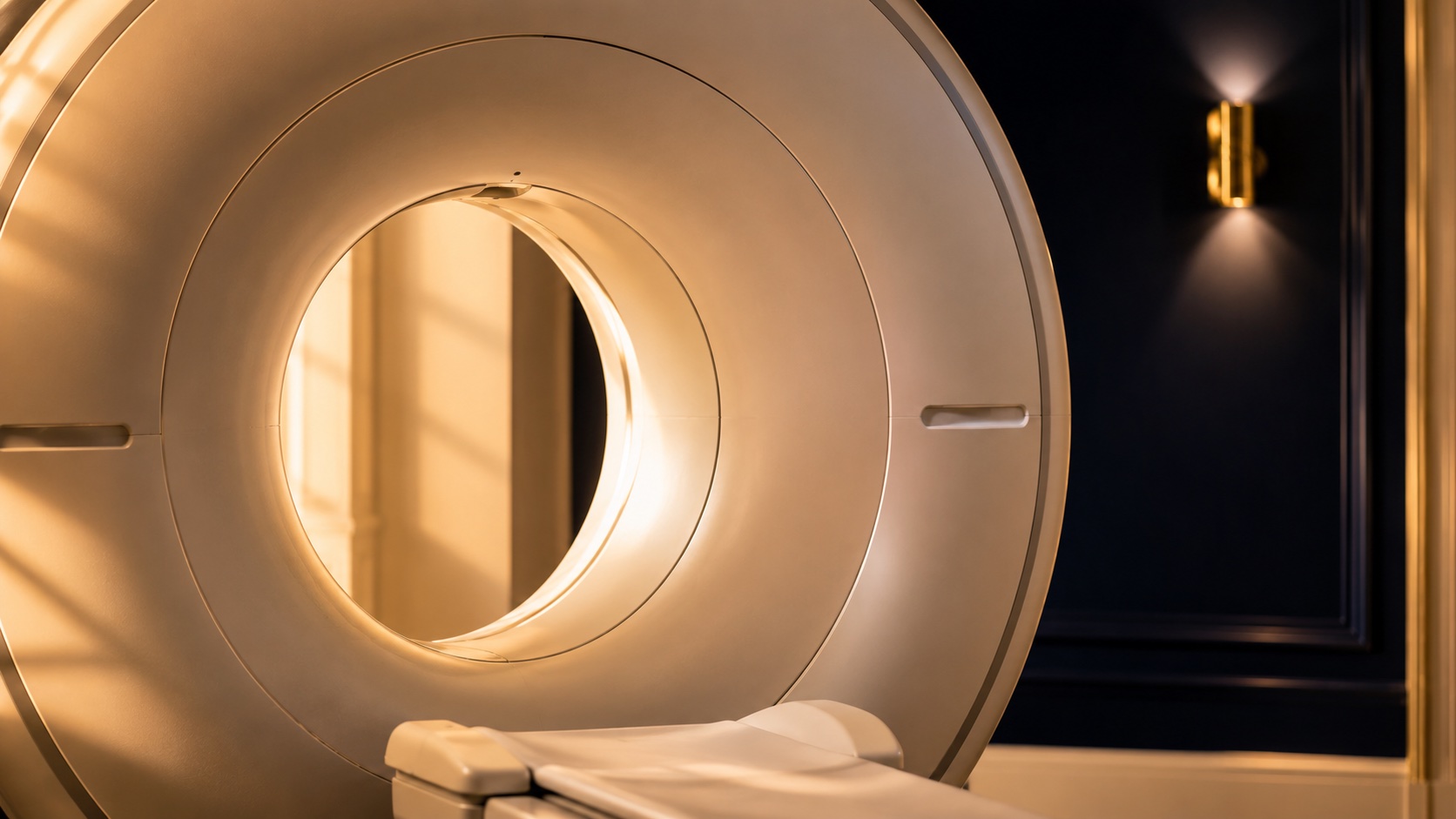
An MRI scanner builds cross-sectional images of the heart from many angles as it beats. There is no radiation. A contrast agent (gadolinium) is sometimes given through a vein to highlight the heart muscle and blood flow.
What does it show?
- The exact size, shape and pumping function of both ventricles
- Scarring from a previous heart attack, and areas of inflammation (myocarditis)
- The heart muscle in conditions such as cardiomyopathy
- Blood flow, and whether muscle can still recover after a heart attack
- The structure of the valves and the great vessels
How does it help with treatment?
Cardiac MRI is often used when the diagnosis is unclear, or when precise measurements will change the plan.
- Confirming or refining a diagnosis after an echo or other tests
- Measuring heart function accurately before decisions about devices or surgery
- Telling scar apart from muscle that can still recover
What happens on the day?
- Before — you remove metal items and tell the team about any implants or a pacemaker
- During — you lie still inside the scanner for 30–60 minutes; it is noisy but painless, and you may be asked to hold your breath briefly; contrast may be given through a small cannula
- After — you return to normal activity; a specialist report follows, usually within a few days