What is a cardiac echo?
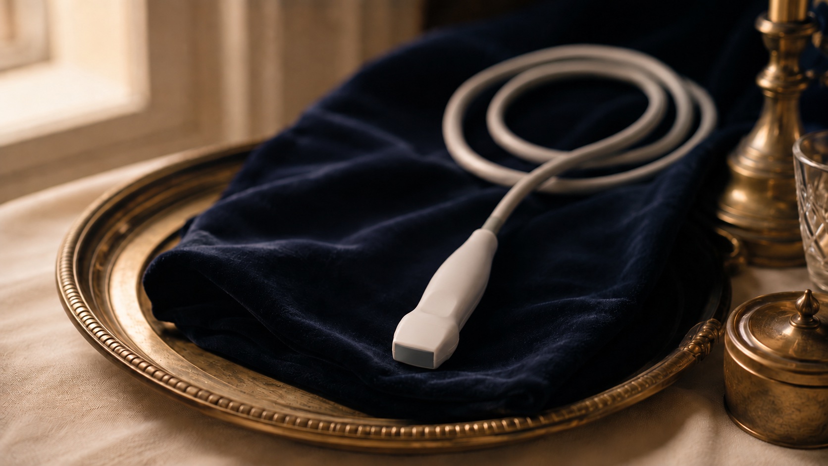
A small probe sends painless sound waves through the chest and builds a live, moving picture of the heart. The most common form is a transthoracic echo (TTE), taken from the surface of the chest. A transoesophageal echo (TOE) uses a thin probe passed into the gullet for a closer view, and a stress echo images the heart during or just after exercise.
What does it show?
A cardiac echo shows the size and structure of the heart, how well it is pumping, and how the valves open and close.
- How strongly the heart muscle contracts
- Whether any valve is narrowed or leaking
- Fluid around the heart, or a clot inside it
- Signs of heart failure or of a previous infection
- How the heart responds to exertion (stress echo)
How does it help with treatment?
Because it is quick, safe and repeatable, an echo is used both to investigate symptoms and to plan care.
- Assessing symptoms such as breathlessness, palpitations or a murmur
- Planning valve repair or replacement, including minimally invasive procedures
- Guiding treatment with live imaging and following it up over time
What happens on the day?
- Before — no fasting for a standard TTE; you may be asked not to eat for a few hours before a TOE
- During — you lie on your side, gel is applied, and the probe is moved over the chest; a TOE is done with sedation and a throat-numbing spray
- After — you can usually return to normal activity straight away, and results are typically ready within 24 hours